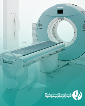
Iraq's first spiral incisive CT scanner at Al Hayat Center
Iraq's first Philips Incisive CT 256 machine, also known as tomography or computed tomography, is an advanced device for imaging the body's layers. X-rays are used in imaging for diagnostic purposes in the medical field. It features advanced technology and a high slice count (256 slices), enabling high-quality images and improving diagnostic accuracy.
The contradiction between optimizing image quality and minimizing radiation dose exposure was an obstacle in conventional CT imaging. Still, AI technologies have overcome these limitations and pushed the spiral incisive CT scanner into another era.
The multi-slice spiral CT scanner has become an important tool in medical diagnosis in several medical specialties such as oncology, cardiovascular diseases, and others. This device allows doctors to see tissues and organs clearly and accurately through high-resolution 3D images, which helps them determine the correct diagnosis and plan the appropriate treatment for patients. It is also effective in taking tissue biopsies from different body areas, such as the lungs, liver, and bones.
The spiral technique in Iraq's first spiral CT scanner:
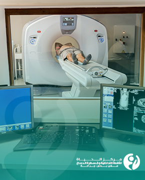
Spiral technology means that the device images the body in a spiral pattern, where the body rotates inside the round ring of the device as the device slowly moves up or down. This continuous movement allows for a series of sequential images of thin layers of the body. This information generates three-dimensional images of the body's internal tissues and structures.
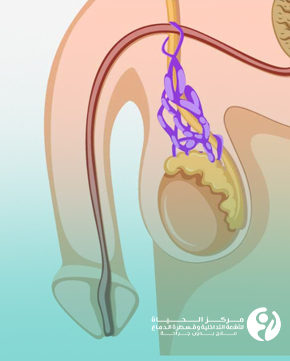
Eclipse Collimator:
The Eclipse Collimator is a beam scattering system used in the Philips Incisive CT. The Collimator accurately directs and focuses the X-rays emitted by the device on the target area for imaging, increasing accuracy and improving image quality. This system minimizes the scattering of rays and focuses them on the desired part, improving resolution and minimizing the noise effect on the final image.
Let's summarise the advantages of the spiral incisive CT scanner at Al Hayat Center for Interventional Radiology and Neurointervention for the first time in Iraq:
•Thanks to the spiral technology and the high number of slices (256 slices), the spiral CT scanner can provide wide body coverage and improve diagnostic accuracy.
•It can deliver accurate and clear three-dimensional images that display fine details of tissues and internal structures such as the brain, heart, lungs, blood vessels, bones, and soft tissues.
•The wide opening and ergonomic design make it easy for patients to get in and out of the device, increasing their comfort during the examination.
•The device is characterized by faster imaging speed and significantly and consistently reduced radiation dose across all patient groups compared to traditional radiology techniques, meaning shorter scan times and fewer potential side effects.
The AI-enabled CT scanner has the ability to:
•Process the CT images within seconds to optimize image quality and minimize image distortion caused by low-dose imaging and the negative impact on imaging quality.
•Comprehensively reducing image distortion while maintaining image quality has a profound impact on the quality and efficiency of imaging procedures, especially for high-risk patients and children.
•Demonstrate subtle changes in diseases.
•Allowing radiologists to interpret images more quickly and accurately; improves the radiology report in terms of accuracy and time spent, unlike the previous situation in which poor quality images caused by high noise were difficult for radiologists to interpret, forcing them to spend more time carefully reviewing, which reduces reading efficiency and increases report preparation time.
•Eliminating concerns about the negative effects of radiation through using the lowest achievable radiation dose.
Overall, the Philips iCT 256 slice / Eclipse Collimator, first introduced in Iraq within the Al Hayat Center for Interventional Radiology and Neurointervention in Karbala is an advanced technology in the field of medical X-ray imaging due to a comprehensive set of AI capabilities, providing high-resolution three-dimensional images and improved diagnosis, with shorter scan times and lower radiation dose.